Ping Meng1 ![]() ,
Shouguo Ma1,
Shuwen Huang2
,
Shouguo Ma1,
Shuwen Huang2
For correspondence:- Ping Meng Email: huangshuwen133494@163.com Tel:+8653183251203
Received: 2 June 2015 Accepted: 5 March 2016 Published: 30 April 2016
Citation: Meng P, Ma S, Huang S. Healing effect of Terminalia chebula Retz extract on second-degree burns in rats. Trop J Pharm Res 2016; 15(4):787-791 doi: 10.4314/tjpr.v15i4.17
© 2016 The authors.
This is an Open Access article that uses a funding model which does not charge readers or their institutions for access and distributed under the terms of the Creative Commons Attribution License (http://creativecommons.org/licenses/by/4.0) and the Budapest Open Access Initiative (http://www.budapestopenaccessinitiative.org/read), which permit unrestricted use, distribution, and reproduction in any medium, provided the original work is properly credited..
Purpose: To investigate the healing effect of Terminalia chebula Retz. Extract (TCRE) on second-degree burns in rats.
Methods: Male Sprague Dawley (SD) rats, weighing 200 – 220 g, were subjected to deep second-degree skin burns by electrical scald instrument. The animals were divided into three groups as follows: (1) second-degree burns model (control) group, (2) burns model treated with 1 % silver sulfadiazine (SSD) group, and (3) burns model treated with 100 mg·mL-1 TCRE group. On days 3, 7 and 14 following the administration of the drug/extract, the wound area and histopathological changes in rat epidermis were evaluated for the various groups. The minimum inhibitory concentration (MIC) of TCRE on Staphyloccocus aureus, Pseudomonas aeruginosa and Escherichia coli were also assessed separately.
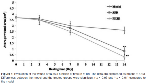
Results: On day 14, the mean wound area of TCRE treatment group (0.25 ± 0.06 cm2) was significantly smaller than that of the control rats (2.71 ± 0.20 cm2, p < 0.01). The histological results indicate that the inflammatory cells disappeared and were replaced by new granulation tissue in the group treated with 100 mg·mL-1 TCRE by day 14. Compared with SSD group rats, the inflammatory cells and fibroblast and granulation tissues of burnt rats treated with 100 mg·mL-1 TCRE were same as those of rats that had no burns. The antibacterial results revealed that the MIC of TCRE on Staphyloccocus aureus, Pseudomonas aeruginosa and Escherichia coli was 3.13, 12.5 and 6.25 mg·mL-1, respectively.
Conclusion: Terminalia chebula Retz. has potentials to be developed as an effective medicinal herb for the treatment of second-degree burns.
Introduction
Every year, millions of people suffer major disabilities or even death from burns, caused by hot water, flame and boiling oil. People suffer from burns due to domestic and industrial accidents, which along with enormous cost of treatment, cause mortality and considerable morbidity [1]. According to the World Health Organization (WHO), there were 300,000 deaths worldwide due to burns in 2012, with 96 % of these deaths occurring in developing countries [2]. Burns are one of the health problems in modern societies associated with irreparable damage to patients and family relationships [3].
Currently, SSD is the most used topical treatment for burns due to its potent antimicrobial efficacy. However, it was found that silver gets absorbed systemically, posing problems on prolonged use and systemic complications such as neutropenia, methemoglobinemia and renal toxicity [4]. Therefore, finding more efficient agents with fewer side effects for treatment of burns has always been a concern of researchers.
Terminalia chebula Retz., a traditional Chinese medicinal herb, is widely distributed in southern China. The root of Terminalia chebula Retz. has been used in treatment of inflammation, infection, jaundice, skin burns and hyperlipemia in China and Japan [5]. Several studies have evaluated the antioxidant capacity of Terminalia chebula Retz. extract [6,7] and its anti-inflammatory activities, such as the inhibition of NF-kB [8]. In the present study, the healing effect of Terminalia chebula Retz. extract on deep second-degree burn wounds in rats was investigated.
Methods
Materials
Herbal samples of Terminalia chebula Retz. were collected from Bozhou City, Anhui Province in China in May 2014. Taxonomic identification of the plant was performed by Prof Ku He of Shandong University of Chinese Medicine in China. A voucher specimen (no. TCRE 201405027) was deposited in the Department of Pharmacy, Zhangqiu Municipal People's Hospital, China for future reference.
The powdered sample (20 g) was placed in a round bottom flask with 70 % ethanol (1: 8, w/v) for reflux extraction at 80 oC. It was extracted twice, 2 h for each, and the ethanol solvent removed with a rotary evaporator, and the remaining solution was concentrated into 200 mL volume as the experimental drug. The working concentration of TCRE was equivalent to 100 mg·mL-1 (extract weights/final volume).
Animals and model preparation
Male SD rats weighing 200-220 g were obtained from Shandong Center for Disease Control and Prevention, Jinan, Shandong. The rats had free access to feeds which were purchased from Guangxi Jiangda Feed Co. Ltd, China, and were allowed to acclimatize for at least one week before use. The animal experiment was approved by the Animal Care and Use Committee of Zhangqiu Municipal People's Hospital (approval reference no. 20131004) and was carried out in compliance with the Animal Welfare Act and the NIH guidelines (NIH publication No. 80-23, revised 1996).
Ethyl carbamate solution (20 %) was used for anesthetizing the rats after the rat hair was shaved off. The top of electrical scald instrument (Changhai Hospital of Second Military Medical University, China) was pressed upon the back skin with a certain force for 15 s at 75 oC. In this way, the second-degree burn model was prepared.
Experimental groups and treatments
All rats were randomly divided into three groups of thirty-five rats each: second-degree burn model group, SSD-treated group and TCRE-treated group. All the animals in the second-degree burn model, SSD-treated and TCRE-treated groups received the deep second-degree burn. SSD cream (1 %, w/w) was used as the standard drug.
In a preliminary study, the dose-response properties of TCRE and silver sulfadiazine were examined to determine the optimal dose, and the most effective in the wound healing was 0.3 g SSD or 1 mL TCRE per wound (data not shown). One mL of TCRE or 0.3 g of SSD was applied slowly with cotton bud to the burn wound area and extended slightly outside the wound area to ensure inclusion of the wound edges. The treatments were repeated twice daily for 14 days. The first application was done directly after the injury. The control group did not receive any treatment for 14 days. The wound remained exposed after the treatment.
Measurement of mean wound area
On days 3, 7 and 14, following treatment, average wound areas of the control, SSD and TCRE group rats were measured. The wounds were photographed with a digital camera in order to calculate the wound surface areas (WSA) with Autocad software 2013 (Autodesk Co. Ltd.). The change in wound surface area in a given day (WSAday-x) was expressed as a percentage of the wound area on the second day (WSAday-2) using Eq 1.
WSA = {(WSAday-2 – WSAday-x)/WSAday-2}100 ……. (1)
Histological studies
Wound skin tissue samples were taken, using a scalpel, from the control, SSD and TCRE groups on days 7 and 14 for histological observation. The skin tissues were fixed with 10 % formalin. After fixation, the samples were embedded in paraffin, cut into 3 mm frozen sections with a cryostat microtome, and then stained with hematoxylin eosin reagent. The collagen fiber, inflammatory cell, blood vessel and granulation tissue of the skin tissues were examined under a microscope.
Antibacterial test
Agar dilution was used to determine the minimum inhibitory concentrations (MIC) of TCRE on Escherichia coli (ATCC23276), Staphyloccocus aureus (ATCC26542) and Pseudomonas aeruginosa (ATCC25338). The three bacteria were all diluted to 1.5 × 10⁵ CFU·mL-1 with 0.9 % sodium chloride solution. The experiment was repeated three times to determine the MIC.
Statistical analysis
The data are expressed as mean ± SEM. Multiple group comparisons were performed using one-way analysis of variance (ANOVA) with SPSS 18.0 software followed by a Dunnett’s test to detect intergroup differences. P < 0.05 was considered statistically significant.
Results
Wound healing
The wound area of burnt rats minished progressively when applied with SSD or 100 mg·mL-1 TCRE treatment. The wounds treated with TCRE healed more quickly than those of the control group. The average wound area of TCRE and SSD treatment groups healed more quickly than the control groups by day 14 (p < 0.01) (), suggesting that both treatment probably accelerated the process of wound healing, hence, there was a significant reduction in wound area. Meanwhile, the mean wound area of burnt rats treated with SSD was significantly smaller than that of the model rats on day 14 (p < 0.01).
Histological features
After the initial burning and elimination of necrotic tissue, a second-degree burn showing muscular and adipose tissues was formed, of which there were neither dermis nor epidermis and infection was absent. Histological findings of the wounded skin, treated with SSD and TCRE on day 7 and 14 are shown in Plate 1. On day 0, collagen fiber was necrotic, inflammatory cells infiltrated below the striated muscles and vascular engorgement and necrosis were seen in the burn skin of model rats. On day 7, there was severe infiltration of inflammatory cells and some fibroblast and granulation tissues were found in the burnt skin of the control rats. The burnt skin of rats treated with SSD or TCRE showed inflammatory cells decreased significantly and fibroblast and granulation tissues grew rapidly. By day 14, inflammatory cells were still seen and some fibroblast and granulation tissues grew in the burnt skin of the control rats. The inflammatory cells disappeared and new granulation tissue, collagen fibers and epithelialization progressed very quickly in rats treated with SSD or TCRE.
Antibacterial test
The MIC of TCRE on Staphyloccocus aureus, Pseudomonas aeruginosa and Escherichia coli were 4.8, 19.2 and 9.6 mg·mL-1, respectively.
Discussion
Thermal burn injury is still a major cause of death and disability in the world and its healing process is a challenge in modern medicine. Burns on human body may be treated by different methods depending on the extent and severity of the burn. SSD is bactericidal on a wide variety of bacteria, so it is commonly used to prevent and treat infections of second and third degree burns. Recent studies revealed that SSD ointment has positive effects on the proliferation of fibroblasts which are the main source of collagen and fibronectin [9]. However, current reports suggest that silver-based products show side effects and researchers are making efforts to seek for better topical antimicrobial products [10].
During the treatment of burn, the key method is to control bacterial infection. The common and main bacteria isolated from clinical burn patients were Staphylococcus aureus, Pseudomonas aeruginosa and Escherichia coli. SSD is the most used topical treatment for burn injury due to its anti-microbial efficacy. However, it has systemic complications such as neutropenia, methaemoglobinemia and renal toxicity.
Traditional Chinese medicine has the advantage of inhibiting bacterial growth with few side effects. It was reported that gallic acid was one constituent of Terminalia chebula Retz, and showed strong antibacterial activity [11]. Therefore, TCRE inhibited the bacterial growth of the burn wound effectively.
In recent years, there has been a growing interest in alternative medicines and natural medicinal products for the local treatment of wounds due to the high costs of traditional drug treatments [12]. Skin integrity is restored by a physiological process aimed at repairing the damaged tissues. The healing process proceeds in four phases: hemostasis, inflammation, proliferation and remodeling [13]. The time required for complete healing of deep second-degree burns, without the application of specific therapeutic agents, can be three to six weeks or more and these burns will leave a scar tissue that may undergo hypertrophy and contract itself [14]. In our study, TCRE accelerated the scab of deep-degree burn wound and prevented infection effectively.
Burn wounds treated with TCRE recovered well by day 14. However, the wounds of the control group still showed severe inflammatory cell infiltration, but the epidermis of burn was healed.
Conclusion
These results reveal that Terminalia chebula Retz can accelerate the healing of second-degree burns and has a strong antibacterial activity. The plant can potentially be developed into a product for burns treatment.
References
Archives


News Updates